
Fig. 2.
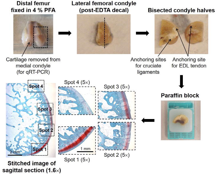
The lateral femoral condyles of rabbit distal femora were decalcified in EDTA, bisected and the halves embedded in paraffin. Sagittal sections of the condyle halves were stained with safranin O/fast green and haematoxylin. Stitched images of the whole surface, as well as four higher-magnification images, were scored according to the system of Laverty et al. (2010): the stitched images were used to score GAG staining and surface structure, while the higher magnification images were used to determine chondrocyte density and clustering.