
Figure 3. Liver ablation zone features on TUNEL staining.
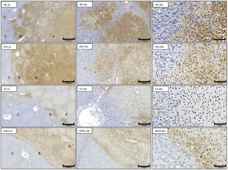
Microscopic appearance at day 1 of IRE, RFA, CA and MW ablation zone with area of necrosis (A), margins (B), and normal liver (C). (TUNEL stain; x2) and transition zone ( x10, x40 magnification).