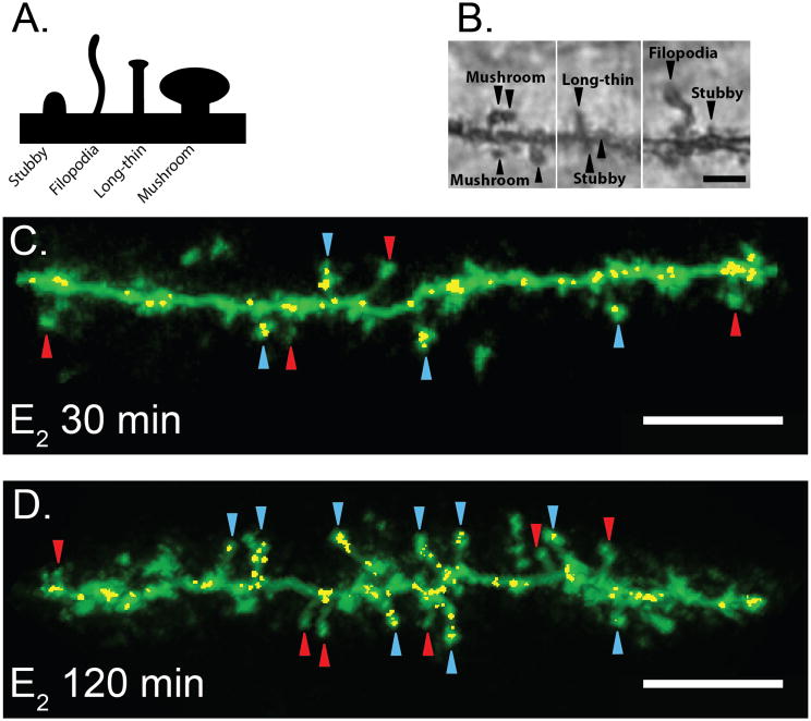
Figure 5. Identification of spine-specific changes in the CA1 region of the hippocampus following Estradiol administration.
(A) Classification of dendritic spine morphology. (B) Representative Golgi-impregnation of hippocampus CA1 spines captured at 100x magnification. Scale bar =3μm. (C, D) Estradiol increases colocalization of GluA2 and PSD95 within Mushroom spines. Representative dendrites from E30 min (C) and E120 min (D). Yellow voxels indicate colocalized GluA2 and PSD95 within Mushroom spines. All carats indicate mushroom spines. Red carats indicate mushroom spines that do not contain colocalized GluA2 and PSD95. Blue carats indicate mushroom spines that contain colocalized GluA2 and PSD95. Scale bar =5μm.

