Figure 3.
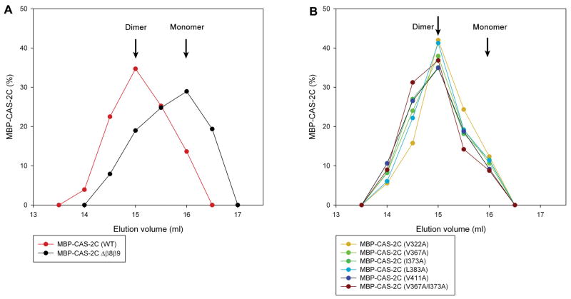
Estimation of dimeric states of MBP-CAS-2C variants by analytical gel filtration chromatography. (A) MBP-CAS-2C (WT) and MBP-CAS-2C Δβ8β9 (lacking the dimerization motif) were applied to Superose 6 gel filtration chromatography (24 ml bed), and eluted fractions were analyzed by SDS-PAGE. Relative amounts of eluted proteins are plotted on the graph. The graph shows the data in the elution volumes of 13 – 17.5 ml, and no other peaks were detected outside of this range. Peak positions of dimeric MBP-CAS-2C (WT) and monomeric MBP-CAS-2C Δβ8β9 are indicated. (B) Elution profiles of MBP-CAS-2C mutants were similar to that of dimeric MBP-CAS-2C (WT), suggesting that dimeric states were preserved in the examined mutants. No other peaks were detected outside of the range of 13 – 17.5 ml.

