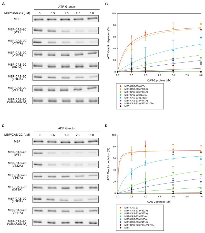
Figure 4.
Binding of CAS-2C variants to ATP- and ADP-G-actin analyzed by supernatant-depletion assays. DyLight 680-labeled ATP-G-actin (0.2 μM) (A and B) or ADP-G-actin (0.2 μM) (C and D) was incubated with various concentrations of MBP or MBP-CAS-2C variants for 1 hr. The MBP-fusion proteins and associated G-actin were pulled down by Ni-NTA resins. DyLight 680-actin in the supernatants was analyzed by SDS-PAGE and infrared imaging (A and C). Percentages of G-actin depletion from the supernatants are plotted as a function of total concentrations of MBP-fusion proteins (B and D). Data are means ± standard deviations (n = 3).

