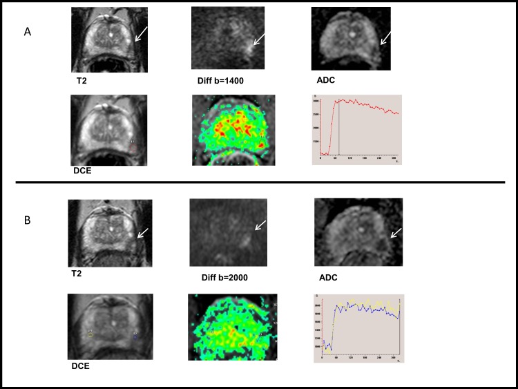
Fig 4. An example of tumour progression.
(A) Prostate MRI taken in September 2013. 71-year-old patient in AS. T2WI shows a small low signal intensity lesion (max diameter 9 mm). Restricted diffusion and focal enhancement was found in left midgland. Routine biopsies revealed Gleason score (GS) 3+3 prostate cancer in left apex and right base. (B) Same patient, prostate MRI taken in January 2015. The lesion appeared larger (max diameter 13 mm) in T2WI and more visible in diffusion and ADC map, while DCE remained unspecific. GS 4+5 prostate cancer was found in cognitively targeted biopsies. Radical prostatectomy in June 2015 proved GS 4+3 disease with two small foci in the right lobe as well. Also, 0.5 mm extraprostatic extension with 5 mm contact to external tissues was diagnosed. Tumour stage was pT3a.

