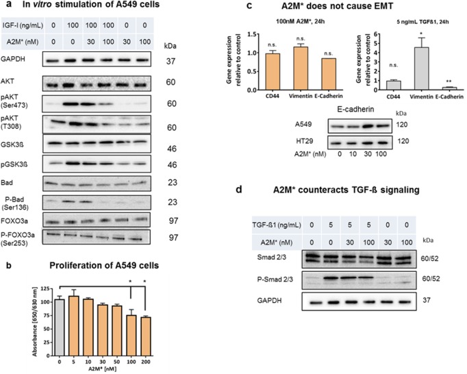
Fig 4. Impact of A2M* at signalling pathway members and EMT in tumour cells.
(a) A549 cells were cultured in the absence (control) or presence of IGF-1 (100 nM) and/or A2M* (30 and 100 nM) for 8h. Cells lysates were analysed by immunoblotting for detection of non-phosphorylated and phosphorylated proteins using antibodies as in shown in (S6 Table). (b) Inhibition of proliferation of A549 cells by A2M* analysed by WST-1 (n = 3), error bars are mean ± sem, t-test. (c) A2M* does not induce epithelial-mesenchymal transition (EMT). A549 cells were cultured in the presence of A2M* (100 nM) or TGF-ß1 (5 nM) for 24h followed by gene expression analysis by qRT-PCR of CD44, vimentin and E-cadherin (n = 4), error bars are mean ± sem, t-test). Immunoblot of E-cadherin in A549 and HT29 cells stimulated with A2M* (0, 10, 30, 100 nM) for 24h normalized to GAPDH. (d) A2M* counteracts TGF-ß1 signalling in A549 cells by inhibition of SMAD phosphorylation. All experiments were performed in triplicates. (*P < 0.05, **P < 0.01).

