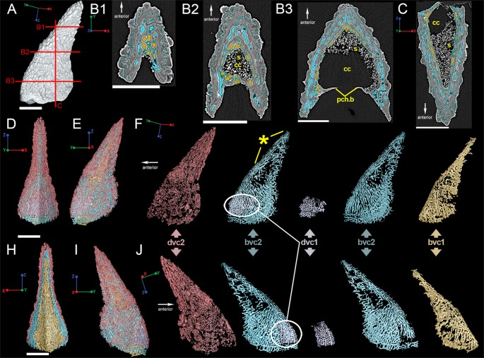
Fig 3. Vascularization of the Romundina dorsal ridge spine using low-resolution synchrotron data (7.46 μm).
(A) Reconstruction of the dorsal spine showing approximate locations of slices B1-B3 and C. Scale bar is 2000 μm. Virtual thin sections taken through the spine on (B1-B3) transverse and (C) frontal planes. Scale bars 1500 μm. (D) Anterior and (E) left lateral views of the reconstructed vascularization in the spine, while (F) deconstructs the vascular canals into layers beginning with the outermost layer in red. These colors relate to the virtual thin sections in (B1-B3). Star indicates relative position of the spine's growth origin. (H) Posterior and (I) right lateral views of the spine with similar vascular break down to (F) but in right lateral view. Scale bars are 2000 μm. Abbreviations are: bvc, bone vascular canals; cc, central cavity; dvc, dentine vascular canals; pch.b, perichondral bone; s, sediment.

