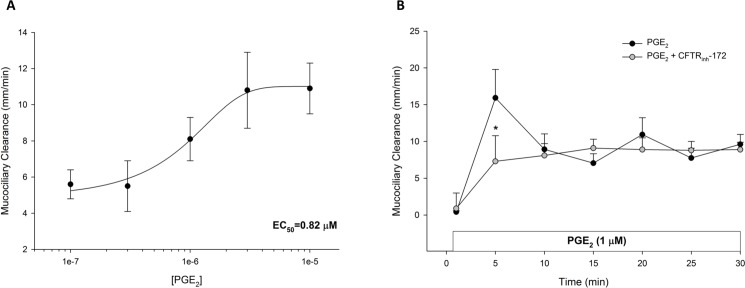
Fig 1. PGE2-stimulated mucociliary transport in ferret trachea.
A. PGE2 stimulates a dose-dependent increase in MCC in ferret trachea. Each tissue was exposed to 2–3 doses of PGE2 for 30 minutes each (n = 3 each dose). Data are shown as the mean PGE2-stimulated increase in MCC over baseline ± SEM. The half-maximal effective concentration (EC50) is noted in lower right corner. B. Timecourse of PGE2-stimulated MCC with and without CFTR inhibition (n ≥ 6 each). For CFTR inhibition, tissues were bathed in apical and serosal solution for 30 minutes with CFTRinh-172 (20 μM) prior to the 15-minute period and kept in the serosal bath for the length of the experiment. PGE2 (1 μM) was added to the serosal bath. Circles represent means with bars indicating SEM. Asterisks represent P < 0.05 by ANOVA.

