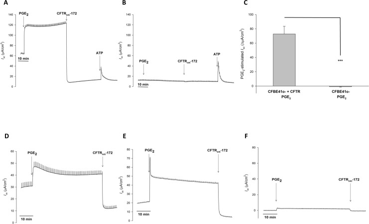
Fig 3. In human bronchial epithelial cells, PGE2 stimulated Cl- secretion is completely CFTR dependent.
A. Representative Isc trace with vertical deflections indicating the change in Isc after a 1 mV pulse was applied (every 1 minute). Bronchial epithelial cells were exposed to serosal to mucosal Cl- gradient with equivalent bilateral HCO3-. PGE2 (1 μM, serosal) was added to HBE41 WT cells after a baseline period of ≥ 10 minutes, with CFTRinh-172 (20 μM, mucosal) added afterwards. To verify cell viability, ATP (500 μM, mucosal) was added. B. Representative Isc trace from a similar experiment with CFBE41 CF cells. C. Change in PGE2-stimulated Isc (mean ± SEM, n ≥ 4) in CFBE41 WT and CF cells. Asterisks denote significance by Student’s t-test (***, P < 0.001). Mean percent inhibition compared to CFBE41 WT noted. D-F. PGE2 stimulated Cl- secretion in 16HBE14o- cells (D), primary cultures of human bronchial epithelial cells (E), and primary cultures from CF nasal polyp extract (F). Experiments were performed in the same manner as Fig 3A and representative Isc traces are shown. N ≥ 3 experiments were performed for each set of cells with similar responses.

