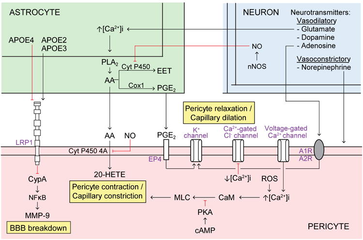
Figure 5. Pericyte-astrocyte and pericyte-neuron signaling pathways.

Astrocytes (top left, green) secrete apolipoprotein E (APOE) 2 and 3 that bind to pericyte lipoprotein LRP1 receptor (bottom, orange) to inhibit downstream CypA-NFκB-MMP-9 pathway. In contrast, APOE4 binds weakly to LRP1, which activates the pro-inflammatory CypA-NFκB-MMP-9 cascade leading to blood-brain barrier (BBB) breakdown. Astrocyte intracellular Ca2+ concentration ([Ca2+]i) increases in response to neuronal factors, for example glutamate, which promotes phospholipase A2 (PLA2)-mediated arachidonic acid (AA) generation. In astrocytes, AA is metabolized into prostaglandin E2 PGE2) via cyclooxtgenase-1 (Cox1), as well as into epoxyeicosotrienoic acids (EET) via cytochrome P450. Astrocytic AA is metabolized into 20-HETE in mural cells via membrane-bound cytochrome P450 4A, which promotes pericyte contraction. PGE2 from astrocytes binds to pericyte EP4 receptor, which alters K+ conductance and promotes pericyte relaxation. Nitric oxide (NO) generated by neurons inhibits cytochrome P450 in astrocytes and cytochrome P450 4A in pericytes to prevent AA to EET and AA to 20-HETE metabolism, respectively. In pericytes, increased cyclic adenosine monophosphate (cAMP) signals via protein kinase A (PKA) to inhibit myosin light chain (MLC) phosphorylation and prevent pericyte contraction. Additionally, pericyte [Ca2+]i increases in response to voltage-gated Ca2+ channels and reactive oxygen species (ROS). Increased [Ca2+]i in pericytes is shown to promote contraction, possibly via downstream signaling through calmodulin (CaM) and MLC kinase (MLCK) which phosphorylates MLC to induce contraction, as shown in VSMCs. Conversely, decreasing [Ca2+]i in pericytes inhibits Ca2+-gated Cl- channels which promotes relaxation. Furthermore, neurotransmitters promote pericyte, relaxation (e.g., glutamate, dopamine, and adenosine) or contraction (e.g., norepinephrine). For example, adenosine signals through adenosine A1 and A2 receptors (A1R, A2R) on pericytes to alter K+ conductance and promote pericyte relaxation.
