Fig. 1.
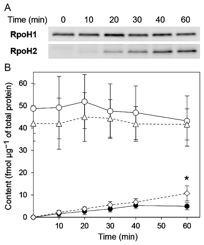
Time course changes in RpoH1 and RpoH2 levels in Sinorhizobium meliloti exposed to heat shock. (A) Representative Western blots. Each lane contains lysates of wild-type cells (1 μg total protein for the RpoH1 analysis and 5 μg for the RpoH2 analysis). Cells were grown in LB medium supplemented with MgCl2 (2.5 mM) and CaCl2 (2.5 mM) at 25°C to an optical density at 660 nm of 0.5 (time 0) and then exposed to 37°C for the indicated time. (B) Quantified data from the Western blot analysis. Contents of RpoH1 in wild-type cells (open circles, solid line) and rpoH2 mutant cells (triangles, broken line), and those of RpoH2 in wild-type cells (closed circles, solid line) and rpoH1 mutant cells (diamonds, broken line) were estimated from band intensities. Values are means±SD from at least six measurements. RpoH2 concentrations were significantly different (t-test, * P<0.001) at 60 min between the wild type and rpoH1 mutant.

