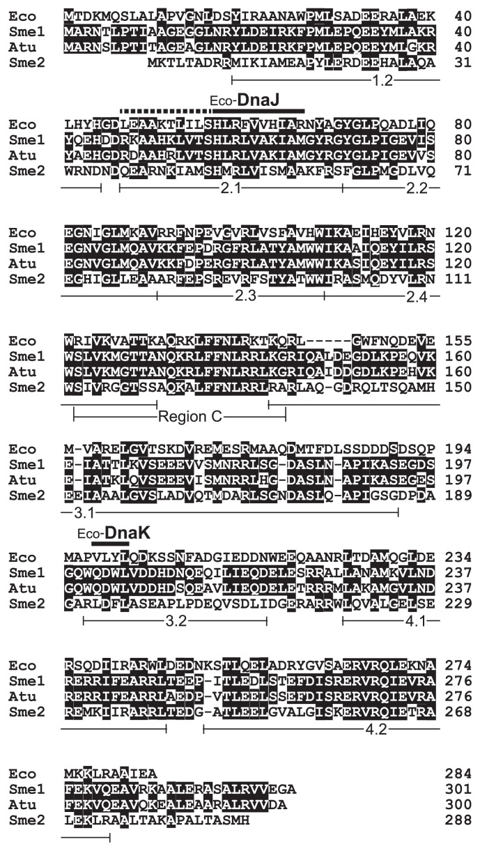
Fig. 2.
Comparison of amino acid sequences of RpoH-type sigma factors. Eco, Escherichia coli σ32 (RefSeq accession number NP_417918); Sme1, Sinorhizobium meliloti RpoH1 (NP_386832); Atu, Agrobacterium tumefaciens RpoH (WP_035228050); and Sme2, S. meliloti RpoH2 (NP_387362). Identical residues at each position are shown in white letters on a black background. The DnaJ- and DnaK-binding sites reported for E. coli σ32 (25) are marked above the alignment; a broken bar (DnaJ) indicates an extension of the binding site proposed later (31). Leu-47, Ala-50, and Ile-54 of σ32 may contact the degradation machinery (21). Regions that are highly conserved among all σ70-family proteins (regions 1.2 to 4.2) (10) and region C, which is unique to RpoH sigma factors (15), are indicated below the alignment.

