. 2017 Dec 14;6:e29750. doi: 10.7554/eLife.29750

Figure 2. cAMP increases the intracellular labile Fe(II) pool in cells.

(A) cAMP (1–100 μM) treatment for 4 hr increased the intracellular labile Fe(II) pool detected by Trx-Puro ferrous iron probes. (B) IF quantification shows the dose-dependent effect of cAMP on labile Fe(II). (C) cAMP (100 μM) treatment for 2–24 hr increased labile Fe(II). (D) IF quantification shows the peak effect of cAMP on labile Fe(II) after treatment for 4 hr. Scale bar = 20 μm. *p<0.0005 (n = 3 independent experiments with three biological replicates in each group, error bars denote standard deviation).

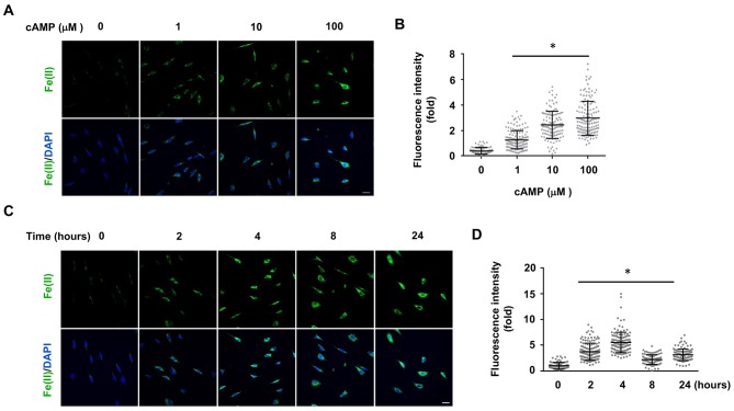
Figure 2.

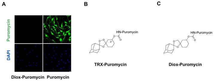
Figure 2—figure supplement 1. Negative control for puromycin incorporation with labile Fe(II) probe TRX-puro in Schwann cells.

Figure 2—figure supplement 1.

(A) No immunofluorescence (IF) signal was observed after incubation with negative control dioxolane compound, Diox-Puromycin, (1 μM) for 2 hr, indicating no puromycin incorporation. Strong IF signal showed after incubating cells with unconjugated puromycin in rat Schwann cells. Scale bar = 20 μm. (B) The chemical structure of TRX-puromycin labile Fe(II) probe. (C) The chemical structure of nonperoxidic dioxolane compound, Diox-Puromycin. The α-amine of puromycin is carbamoylated in both compounds. Consequently, these two conjugated compounds cannot be incorporated into nascent polypeptides, unlike unconjugated puromycin. Labile Fe(II) can react with the 1,2,4-trioxolane ring of TRX-Puromycin causing release of the puromycin and its incorporation into nascent polypeptides, which can then be detected by anti-puromycin antibody. Labile Fe(II) does not react with the bioisosteric nonperoxidic dioxolane compound, Diox-Puromycin, which was used as negative control for all labile Fe(II) IF experiments (n = 6 independent experiments with three biological replicates).
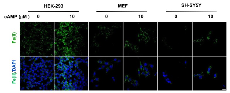
Figure 2—figure supplement 2. cAMP increases the intracellular labile Fe(II) pool in different cell types.

Figure 2—figure supplement 2.

cAMP (10 μM) also enhanced the intracellular labile Fe(II) pool detected by Trx-Puro probes in HEK-293, MEF, and SH-SY5Y cells after treatment for 4 hr. Scale bar = 20 μm.
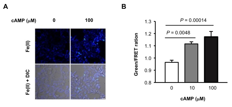
Figure 2—figure supplement 3. (A) Representative ratiometric confocal microscopy images of live HEK-293 cells loaded with FIP-1.

Figure 2—figure supplement 3.

Scale bar = 20 μm. (B) Quantification of mean Green/FRET ratios, which represent the relative abundance of labile Fe(II) in the cell. Statistical significance was assessed by calculating p-values using one-way ANOVA with the Bonferroni correction.