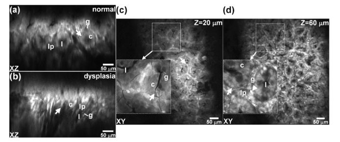
Fig. 11.
Using λex = 561 nm, fluorescence images collected in vivo in the oblique plane from a) normal and b) dysplastic colonic epithelium from mouse colonic epithelium expressing tdTomato are shown. Images collected ex vivo in horizontal plane at depths of Z = c) 20 and d) 60 μm are shown. Electronically magnified ROIs with dimensions of 80 × 80 μm2 (inset) clarify individual cells. Key: crypt (arrow), lumen (l), goblet cells (g), cytoplasm (c), inflammatory cells (arrowhead), lamina propria (lp).

