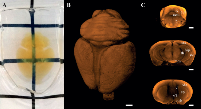
Fig. 3.
Photograph of a mounted and cleared 8-month-old B6SJL F1 mouse brain (A). 3D rendering of the OPT image obtained from tissue autofluorescence (B, Visualization 1 (6.3MB, avi) ). Single coronal sections of the OPT image (C) showing the inner morphology of the intact organ: cc, corpus callosum; cent, central lobule; cp, caudoputamen; ctx, cerebral cortex; cul, culmen; hip, hippocampal region; ic, inferior colliculus; mm, medial mammillary nucleus; och, optic chiasm; p, pons; th, thalamus; v3, third ventricle; vl, lateral ventricle. Grid spacing (in A): 1 cm. Scale bars: 1 mm.

