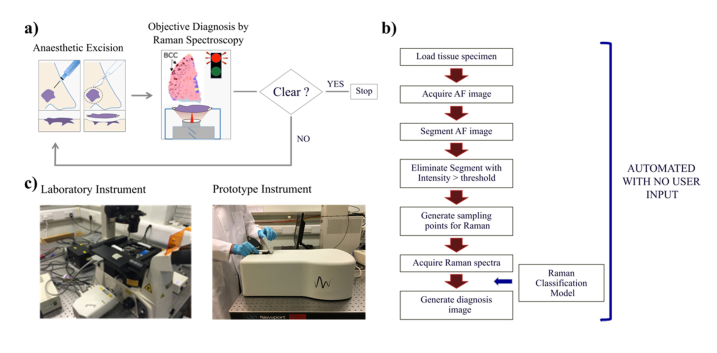
Fig. 1.
a). Schematic description of the intended use of MSH for checking completeness of tumour removal during BCC surgery (Mohs surgery and wide-local excision). b) Flow chart describing the automated measurement and diagnosis algorithms for MSH. c) Photographs of the manual Laboratory Instrument and the automated MSH Prototype. The tissue specimen is loaded in a purpose-built cassette with a quartz bottom window (2.5mm x 2.5cm, 1mm thick). The cassette is manually placed on the microscope stage of the Laboratory Instrument or loaded into the Prototype Instrument.

