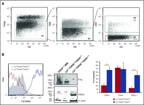
Figure 1.
Differential expression of E-selLs on CD34pos and CD34neg subsets isolated from the Linneg CD38neg fraction of human BM. (A) Overview of the gating strategy used to isolate CD34negCD38neg and CD34posCD38neg fractions by fluorescence-activated cell sorting of lineage-depleted (Linneg) BM MNCs. Left panel, dot plot represents the cell surface expression of a lineage marker cocktail (including CD2, CD3, CD11b, CD14, CD15, CD16, CD19, CD56, CD123, CD235a, and CD7). Cells residing in the negative fraction (R1) were further gated for CD38-negative cells (R2) (middle panel) and then subdivided into 2 subpopulations based on CD34 expression, CD34pos and CD34neg residing in R3 and R4 gates, respectively (right panel). Data shown are representative of n = 4 experiments. (B) Left panel, representative E-Ig staining profile of CD34pos and CD34neg subpopulations isolated as depicted in panel A. The shaded curve shows EDTA control (20 mM; on the LinnegCD38negCD34pos subset), whereas dotted red and solid blue curves show the specific binding of CD34neg and CD34pos subsets, respectively (n = 4). Middle panel, lysates of CD34pos BM cells (CD34pos-BM), LinnegCD38negCD34pos, and LinnegCD38negCD34neg populations isolated from human BM were normalized for total protein level and subjected to western blot analysis. Membranes were blotted with E-Ig, CD44, or β-actin followed by isotype-matched HRP-conjugated mAb for visualization. This is representative of n = 4 independent experiments. Supplemental Figure 1 shows western blots where CD44 was immunoprecipitated from these cell populations and blotted with E-Ig, CD44, and HECA-452. Right panel, flow cytometric analysis of E-selLs expressed on the 2 subpopulations isolated as in panel A is shown as the average percent of expression (above the isotype control) of n = 3 independent experiments. *P < .05 relative to CD34neg subpopulation. NIH Image J was used to quantify the intensity of western blot bands using the gel analyzer tool; the number displayed represents the density of each band related to the LinnegCD38negCD34neg band as a standard. FSC, forward scatter.

