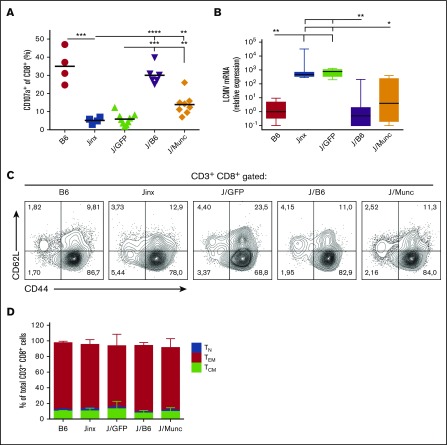
Figure 5.
Functional and phenotypic characterization of CD8+CTLs. (A) To assess degranulation activity, splenic CD8+ T cells were stimulated with murine anti-CD3 (3 µg/mL). Surface expression of CD107a was determined by flow cytometry and presented as percentage of CD8+ population that was CD107a+. P values were calculated using an unpaired Student t test. (B) The viral titer in the liver was measured by a qPCR assay of amplified LCMV mRNA and was normalized against levels of endogenous murine β-actin. The data are presented in a box plot, and P values were determined using a Kruskal-Wallis test for multiple comparisons. Representative fluorescence-activated cell sorter plots (C) and a bar graph (D) showing the distribution of CD8+ T-cell subsets (mean ± standard deviation) at euthanasia (TN: CD62L+CD44−; TCM: CD62L+CD44+; TEM: CD44+CD62L−). *P < .1; **P < .01; ***P < .001; ****P < .0001.

