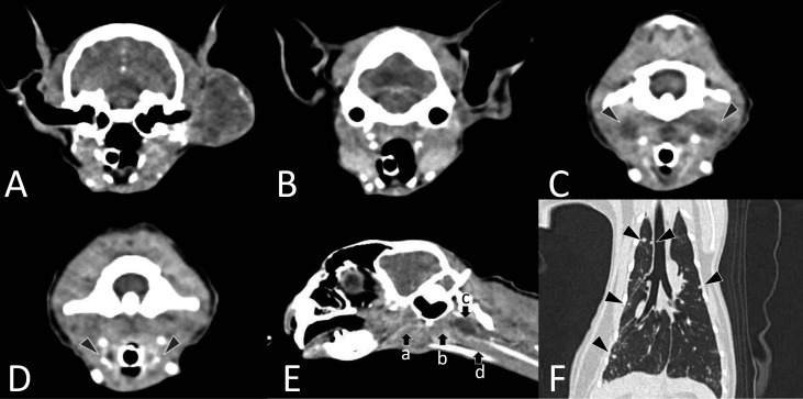
Fig. 2.
Computed Tomography images of the head (2A–E) and thorax (2F). 2A, a soft tissue mass was seen extending around the left ear canal and temporal bone, and it was heterogeneously enhanced by contrast injection. Invasion of the bone tissue was not observed. 2B, image at the level of the mandibular gland and the fore level of the retropharyngeal lymph nodes. Continuity to the periauricular mass is not observed. 2C, medial retropharyngeal lymph nodes (arrowheads) were enlarged and surroundings areas were heterogeneously enhanced by contrast injection. 2D, the laryngeal region (arrowheads) was markedly enhanced by contrast injection. 2E, each position of the mass (a, b), retropharyngeal lymph nodes (c), and laryngeal lesion (d). There are no continuity from periauricular mass to laryngeal lesion. 2F, multiple nodules (arrowheads) were detected throughout the lung lobes.

