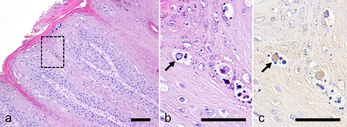
Fig. 1.
Histopathological and immunohistochemical findings. (a and b) Haematoxylin and eosin stain. (b) a higher magnification of the boxed part of (a). (c) PV immunostaining. The neoplasm consists of papillary growth of squamous epithelium (a). There are intranuclear inclusion bodies (b) that are immunopositive for PV antigen (arrow). Bar=100 µm (a); 50 µm (b and c).

