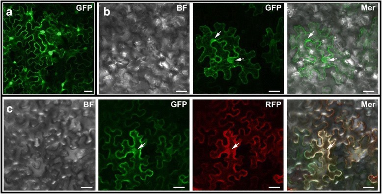
Fig. 6.

Subcellular Localization of OsLAP6/OsPKS1 in tobacco leaf epidermal cells. Confocal images of tobacco leaf epidermal cells after 72 h of infection were shown. a Transient expression of control, showing that the expression of the GFP protein was distributed throughout the cell. b Transient expression of OsLAP6/OsPKS1-GFP, showing that OsLAP6/OsPKS1 may localize to the ER-like structures. c Co-expression of OsLAP6/OsPKS1-GFP and ER-marker, showing the GFP signals of OsLAP6/OsPKS1-GFP are well merged with the RFP signals of ER-marker. The white arrows indicate the ER-ring. BF, bright field; GFP, green fluorescent protein channel; Mer, merged image of each channel. Scale bars = 20 μm
