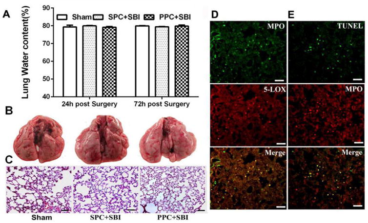
Fig. 3.

PPC induced mild pulmonary inflammation after SBI. (A)Water content of the lungs showed no significant difference within Sham, SPC+SBI and PPC+SBI groups at 24h and 72h after surgery. Data are expressed as mean±SEM. n=6/group. ANOVA, SNK. (B)Representative pictures show gross lung morphology in Sham, SPC+SBI and PPC+SBI groups at 24h after surgery. Gross lung morphology was similar in the three groups at 24h post surgery. n=6/group. (C) H&E staining of lung tissues demonstrated no obvious morphological changes in the three groups at 24h post surgery. Scale bar=100μm. n=2/group. Data not quantified. (D) PPC+SBI group lung samples showed that MPO-positive leukocytes co-localized with 5-LOX and (E) Some MPO-positive leukocytes were TUNEL-positive at 24h post SBI. Scale bar=50μm (D and E). n=2/group. Data not quantified (D and E).
