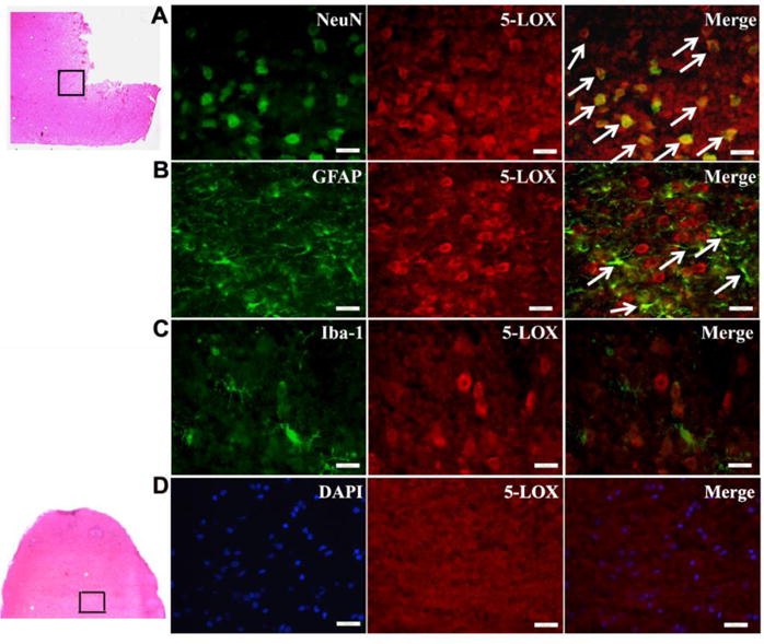
Fig. 6.

Subcellular distribution of 5-LOX in the ipsilateral hemisphere at 24h post SBI. 5-LOX is expressed mainly in NeuN-labeled neurons (A) and GFAP-labeled astrocytes (B) but not in Iba1-labeled microglia (C) 24h after SBI. Arrows indicate colocalization of 5-LOX with NeuN (A) and GFAP with Iba1 (B). Sham control group did not show 5-LOX staining in the right frontal region at 24h (D). Scale bar=25μm(A-C)and 50μm(D). n=2/group. Data not quantified.
