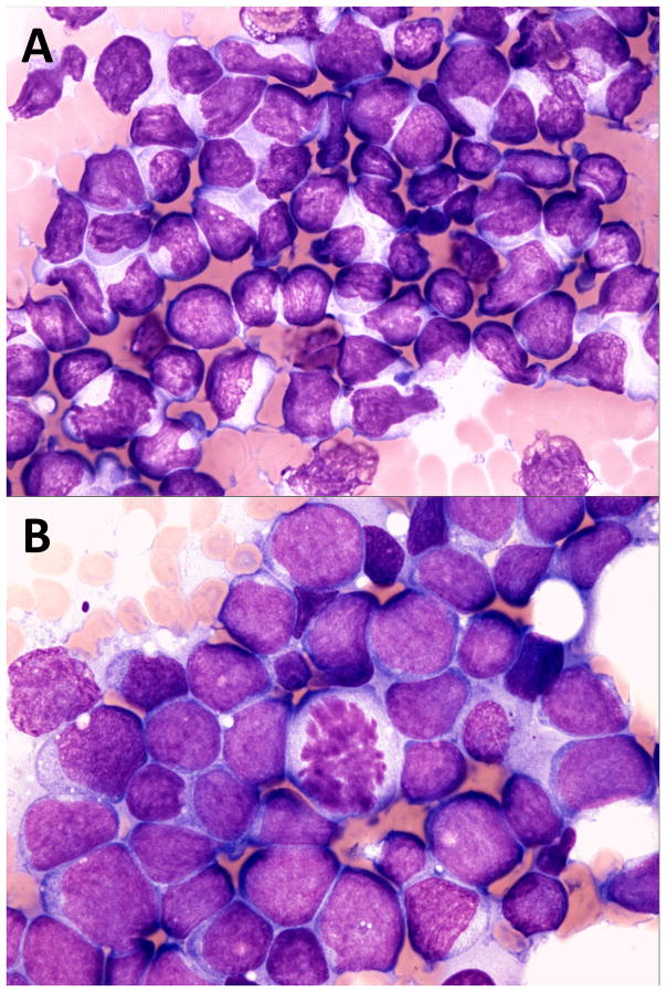
FIGURE 1.
A, Mediastinal lymphoma in a dog, lymphoblastic cell type. Note intermediate size nuclei approximately 1.5–2.0 red blood cells (RBCs) in diameter, immature chromatin with inapparent or inconspicuous nucleoli and frequent nuclear membrane irregularity. ×60 objective magnification, Wright Giemsa stain. B, Mediastinal lymphoma in a dog, large cell type. Note large nuclei approximately 2.5–3.5 RBCs in diameter, immature chromatin and variably prominent nucleoli. ×60 objective magnification, Wright Giemsa stain

