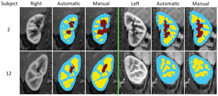
FIG. 7.
The automatic segmentation results of two subjects. On each half, the leftmost column is a selected temporal phase of the DCE-MRI image, the middle column is the output of the automatic segmentation algorithm and the rightmost column is the manual segmentation map (i.e. ground truth). Extended version of this graph can be seen in Supporting Figure S1.

