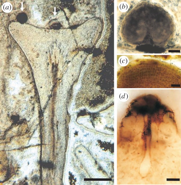
Figure 6.

Fossilized gametangiophore of Lyonophyton rhyniense from the Rhynie chert. (a) Longitudinal section through a cup-shaped gametangiophore bearing two visible antheridia on the upper surface (arrows). The subtending axis contains a central strand of vascular tissues. Scale bar, 1 mm. (b) Longitudinal section through a spherical antheridium. Scale bar, 100 µm. (c) Sperm cells inside antheridium. Scale bar, 30 µm. (d) Archegonium in longitudinal section showing neck, neck canal and egg chamber. Scale bar, 30 µm. Reproduced by permission of the Royal Society of Edinburgh from [62].
