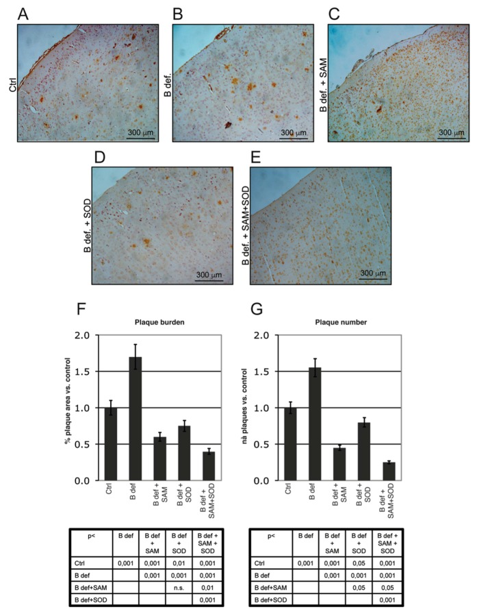
Figure 6.
Amyloid plaque burden in TgCRND8 mice brain. Representative micrographs (A–E) showing amyloid plaques detection in histological sections of the adult mouse brain (Prefrontal Cortex). Magnification 10X. Please note that TgCRND8 mice typically form amyloid plaques at 3 months of age (Ctrl). Plaque area, calculated as % of plaque area/total brain area, (F) and plaque number (G) values of treated mice are expressed taking that of control mice as 1. Statistical significance is listed in the tables below the histograms. Abbreviations as in Figure 3.

