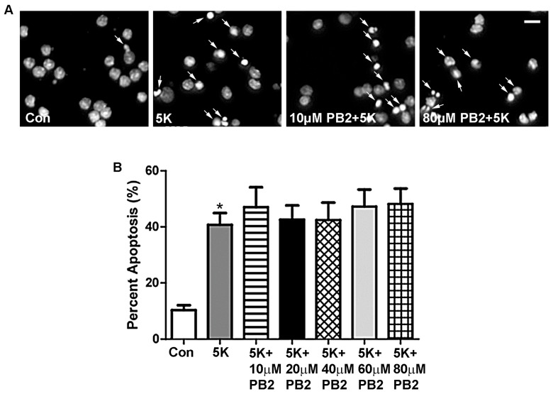
Figure 2.
PB2 does not protect CGNs from 5K-induced apoptosis. (A) Representative images of CGNs treated for 24 h with varying concentrations of PB2 and 5K medium, 5K medium alone, and untreated control cells. Panels show decolorized Hoechst fluorescence to clearly indicate nuclear morphology. Scale bar = 10 μm. Arrows point to cells which were scored as apoptotic. (B) Quantitative analysis of percentage apoptosis of CGNs treated as in (A). Total cells and apoptotic cells were quantified and used to calculate percent apoptosis. Condensed or fragmented nuclei were considered to be apoptotic. Data are expressed as the mean ± the standard error of the mean (SEM), n = 3. Data were analyzed by one-way ANOVA with a post hoc Tukey’s test. * indicates p < 0.05 compared to untreated control. No significant differences were observed between the 5K control and 5K with the addition of any concentration of PB2.

