Figure 6.
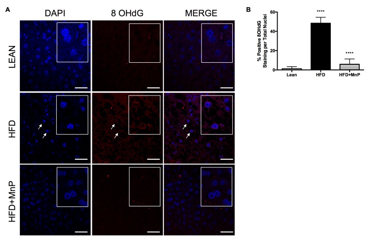
MnP treatment reduces oxidation-induced DNA damage. (A) 8-OHdG immunofluorescence staining of livers after 12 weeks of HFD feeding, with or without MnP treatment. Representative images are shown with a 40× magnification. Arrows indicate hepatocyte nuclei positive for 8-OHdG. The inset boxes show a magnified view of the nuclei; (B) Quantification of 8-OHdG staining. Data are displayed as mean ± SD, n = 3 mice per group. Three images per section were analyzed using Image J Software (National Institute of Health (NIH), Bethesda, MD, USA). Significance was calculated by one-way ANOVA with a Tukey post-test of multiple comparisons. **** indicates p ≤ 0.0001 between Lean and HFD, and **** indicates p ≤ 0.0001 between HFD and HFD + MnP. Images were created in Photoshop (Adobe Systems Inc., San Joes, CA, USA).

