Figure 1.
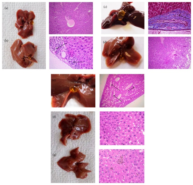
Histopathological microscopic examination in mice liver tissues. Control (a). Hypercholesterolemia (b). Ezetimibe 10 mg/kg (c). ECHE, 100 mg/kg (d). ECHE, 500 mg/kg (e). Hexa-O-acetyl-D-mannitol 10 mg/kg (f). D-Mannitol, 10 mg/kg (g). There were no apparent histological changes in control group fed with a regular chow diet (Figure 1(a)). In Figure 1(b), black arrows indicate the epithelial hyperplasia and granulocyte infiltration in liver of CD1 mice after being fed with a hypercholesterolemic diet. In Figure 1(c) the liver treated with ezetimibe also presented inflammatory infiltration and epithelial hyperplasia; black arrows indicate hepatocellular necrosis. In Figure 1(d) there were no apparent changes after treatment with ECHE 100 mg/kg. In Figure 1(e) all liver tissues showed fibrosis bands (black arrows) and epithelial hyperplasia. There were no apparent histological changes in groups treated with hexa-O-acetyl-D-mannitol (Figure 1(f)) or D-mannitol (Figure 1(g)).

