Fig. 5.

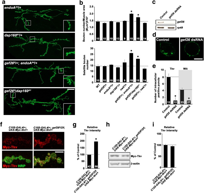
Gef26 regulates endocytic internalization and surface expression of BMP receptors. a and b gef26 interacts with endocytic mutations during synaptic growth. a Confocal images of anti-HRP-labeled NMJ 6/7 in endoA Δ4/+, dap160 Δ1/+, gef26 6/+; endoA Δ4/+, and gef26 6/dap160 Δ1 third-instar larvae. Scale bar, 20 μm. b Quantification of total bouton number and satellite bouton number at NMJ 6/7 in the following genotypes: wild-type, gef26 6/+, endoA Δ4/+, dap160 Δ1/+, nwk 2/+, gef26 6/+; endoA Δ4/+, gef26 6/dap160 Δ1, and gef26 6/+; nwk 2/+. c-e Gef26 is required for endocytic internalization of surface BMP receptors. BG2-c2 cells were transfected with pAc-Myc-tkv-Flag or pAc-Myc-wit-Flag in the absence (control) and presence of gef26 dsRNA. Live control and Gef26-depleted cells were prelabeled with anti-Myc (green) at 4 °C, followed by incubation at 25 °C for 10 min to allow internalization of the labeled surface receptors. After fixation and permeabilization, cells were sequentially stained with anti-Flag (red) and fluorescently-labeled secondary antibodies. c Reverse transcription (RT)-PCR analysis to confirm knockdown efficiency of Gef26. d Single confocal sections through the middle of control and gef26-knockdown cells are shown for the green channel only. Scale bar, 5 μm. e Quantification of the number of intracellular Myc-positive puncta per cell. Only cells with similar Flag signal (red) intensities were analyzed. f and g Steady-state levels of surface Tkv are increased at the NMJ of gef26 mutants. f Representative confocal images of NMJ 6/7 in C155-GAL4/+; UAS-Myc-tkv/+ and C155-GAL4/+; gef26 6 /Df; UAS-Myc-tkv/+ larvae. NMJ preparations were sequentially stained with anti-Myc (red) and anti-HRP (green) under nonpermeant and permeant conditions. Scale bar, 5 μm. g Quantification of the ratio of surface Myc-Tkv to HRP fluorescence intensities. h and i Transgenic expression of Myc-Tkv in neurons is not altered by loss of Gef26. h Western blot of central nervous system (CNS) extracts from C155-GAL4/+; UAS-Myc-tkv/+ and C155-GAL4/+; gef26 6 /Df; UAS-Myc-tkv/+ larvae. The blot was probed with anti-Myc and anti-β-actin. i Quantitative analysis of three independent blots by densitometric measurements. For each sample, the band intensity of Myc-Tkv was normalized to that of β-actin. The number of NMJs (b), cells (e), or synaptic boutons (g) analyzed is indicated in each bar. Data are expressed as mean ± SEM. *P < 0.001
