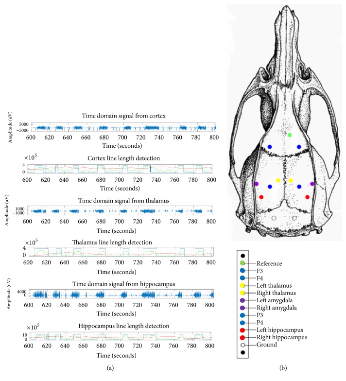
Figure 1.
(a) Field potential recordings from the somatosensory (frontal) cortex, centromedian thalamus, and dentate gyrus of the hippocampus during convulsive seizures. Seizures were detected from corticosubcortical structures using line length. (b) Anatomical targets for implantation of depth electrodes.

