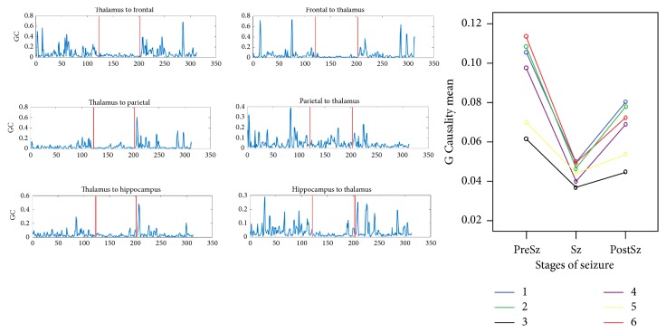
Figure 5.
(a) and (b) represent changes in effective connectivity (EC) during seizure (Sz), preseizure (PreSz), and postseizure (PostSz) in the derivations: (1) thalamus to frontal, (2) frontal to thalamus, (3) thalamus to parietal, (4) parietal to thalamus, (5) thalamus to hippocampus, and (6) hippocampus to thalamus. There was a decrease in the EC from the PreSz to the Sz and then a recovery of the EC in the PostSz state that was lower than the PreSz state.

