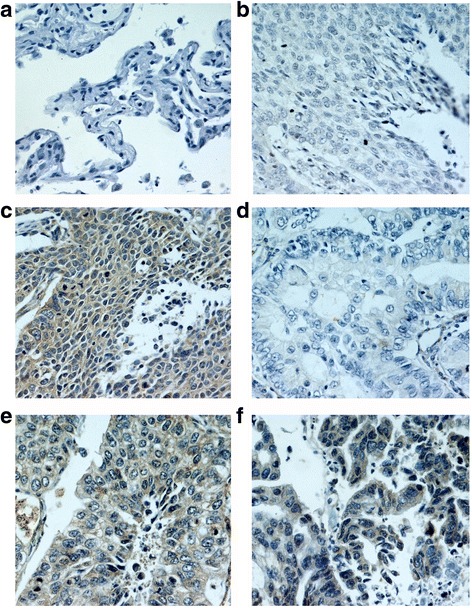
Fig. 1.

IHC analysis of EMMPRIN in NSCLC and normal tissues (IHC×400) a. No staining of EMMPRIN in normal tissues; b. No staining of EMMPRIN in well differentiated LSCC; c. Intensive staining of EMMPRIN in poorly differentiated LSCC; d. No staining of EMMPRIN in well differentiated LAC; e. Moderate staining of EMMPRIN in moderately differentiated LAC; f. Intensive staining of EMMPRIN in poorly differentiated LAC; NSCLC, non-small cell lung cancer; LAC, lung adenocarcinoma; LSCC, lung squamous cell carcinoma
