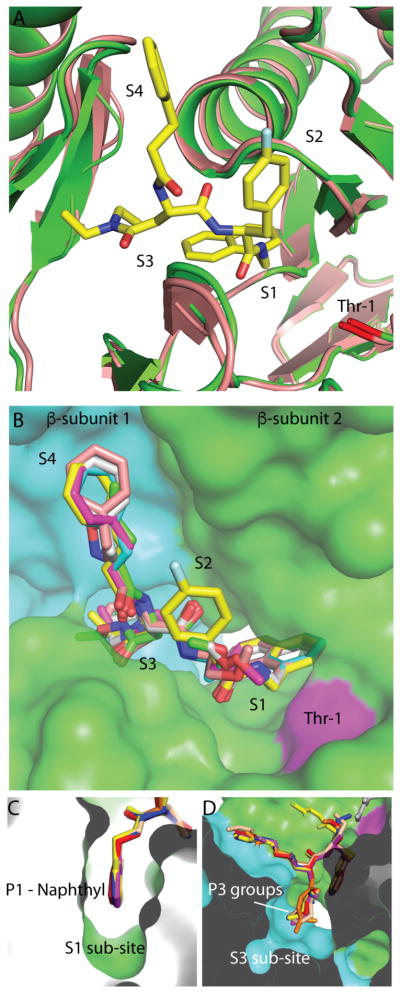
Figure 3. Overview of dipeptide-20S interactions.
(A) Comparison of DPLG-2 bound active site with apo-Mtb20SOG. No significant conformational change was observed in the S1, S2, S3, or S4 sites. DPLG-2 form is in green; apo-form is in wheat. (B) Overview of inhibitor-occupied binding pockets. All six inhibitors are aligned in accordance with the DPLG-2 bound structure. The two β-subunits forming the inhibitor binding sites are shown in green and cyan; Thr-1 is in magenta. (C) The P1 naphthyl groups in the S1 binding pocket. (D) The P3 groups of six inhibitors in the S3 binding pocket.

