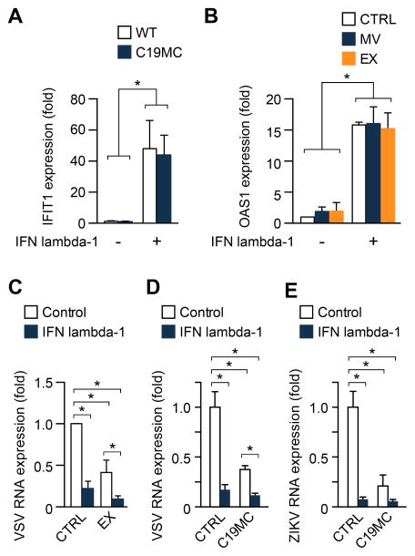
Figure 4. IFN lambda-1 and C19MC miRNAs function independently to confer viral resistance.
(A) The level of the ISG IFIT1 was determined by RT-qPCR in U2OS cells stably expressing the C19MC BAC compared to wild-type, non-transfected cells. Cells were exposed to 10 ng of IFN lambda-1. (B) The level of the ISG OAS1 was determined by RT-qPCR in U2OS cells exposed to microvesicles (MV), or exosomes (EX) with or without the addition of IFN lambda-1 (10 ng). (C) U2OS cells exposed to PHT-derived EX with or without the addition of IFN lambda-1 (10 ng) were infected with VSV at an MOI of 1 for 5 h, and infection was determined by RT-qPCR. Infection level was expressed relative to negative control non-conditioned medium. (D–E) represent the same experiments as in (C), but with C19MC mix (Fig. 1) replacing the EX in cells infected by VSV (D) or ZIKV (E). Experiments were performed three independent times. Differences were determined using ANOVA, with post hoc Bonferroni test. Data are shown as mean ±SD. (*p<0.05).

