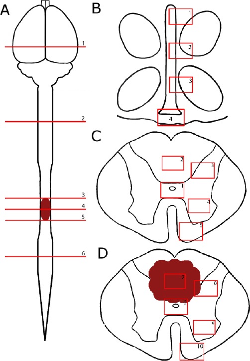
Figure 1.

A schematic of different regions of the neuroaxis being investigated in thoracic spinal cord injury rats.
(A) The levels of the neuroaxis being investigated include: (1) the third ventricle, (2) cervical enlargement, (3) thoracic region 2.25 mm rostral to the epicenter, (4) thoracic region at the epicenter, (5) thoracic region 2.25 mm caudal to the epicenter and (6) lumbar enlargement. (B) Regions of the third ventricle include: (1) alpha region, (2) dorsal region, (3) ventral region, and (4) median eminence. Areas of the (C) normal and (D) injured spinal cord being investigated include (1, 6) central canal, (2) posterior median septum, (3) dorsal grey matter, (4, 9) ventral grey matter, (5, 10) ventral white matter, (7) lesion center and (8) lesion edge in spinal cord injury only.
