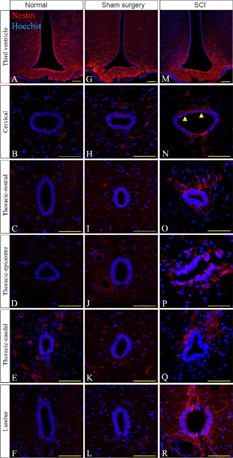
Figure 3.

Immunohistochemical images of nestin reactivity in the ependymal layer in thoracic spinal cord injury (SCI) rats.
(A, G & M) Nestin reactivity (red) was highest at the third ventricle throughout the normal, sham surgery and SCI groups. (N–R) High nestin immunoreactivity was found at all regions of the spinal cord at the central canal in the SCI group compared to the normal (B–F) and sham surgery groups (H–L). For the non-injured cords, equivalent anatomical areas to the injury sites (posterior median septum and dorsal grey matter) were used instead of the lesion center and lesion edge respectively to compare nestin reactivity in each group. Hoechst 33342 solution was used as a counterstain for nuclei (blue). Scale bars: 25 μm. Arrowheads point to nestin positive cells.
