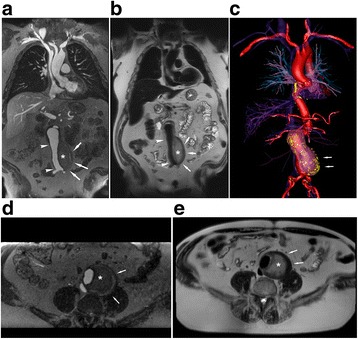
Fig. 5.

Ferumoxytol enhanced (FE) coronal and axial CMR images (3 T) belonging to a 71-year-old male with an infrarenal abdominal aortic aneurysm post endovascular aorto-bifemoral stent repair. Ferumoxytol CMR was performed to evaluate for endoleak in the setting of renal impairment. Coronal and axial bright blood high resolution CMR angiogram (a, d) and FE-HASTE (b, e) images demonstrate a widely patent stent graft (white arrowhead) without endoleak. Note the contrasting intraluminal bright blood signal compared to uniformly dark blood signal suppression in all vascular lumens and intra-cardiac chambers (b, e, Additional file 2: Video S2). Thin aortic valve leaflets are clearly depicted (b). FE-HASTE depicts thrombosis of the aneurysm sac (asterisk, b and e), which is located outside of the endograft. 3D color volume rendered bright blood CMR angiogram (c, Additional file 3: Video S3) illustrates the relationship between the thrombosed aneurysm sac (white arrows) and the endograft
