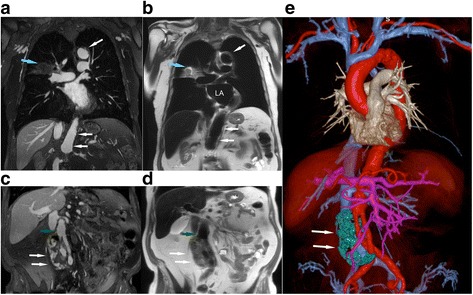
Fig. 6.

Post-ferumoxytol 3D CMR angiogram multiplanar reformat (a, c) and T2 FE-HASTE (b, d) coronal CMR images (3 T) belonging to an 85-year old male with infrarenal abdominal aortic aneurysm post endovascular aorto and bilateral iliac stent graft repair. The CMR exam was performed to evaluate for endoleak. There is diffuse atherosclerosis of the transverse and abdominal aorta with eccentric and ulcerated plaques (a-b, white arrow; Additional file 4: Video S4). An incidental heterogeneous lesion with serpiginous border (a-b, blue arrow) is present in the right middle lobe and is well demarcated on FE-HASTE. Coronal FE-HASTE (b, d) demonstrates uniformly suppressed blood signal throughout the lumen. The aneurysm sac (c-e, white arrows) has circumferential contrast leakage (c-d), suggestive of a Type II endoleak. Relationships between the endoleak (e, white arrow, segmented green) and stent grafts are depicted on the 3D color volume rendered bright blood CMR angiogram image (e). Compared to the bright blood CMR angiogram image (c, green arrow), the intra-luminal blood signal is completely suppressed in the patent iliac stent graft (d, green arrow). Comparison of aortic vessel wall characteristics and endoleak tissue characteristics on FE-HASTE and bright blood 3D CMR angiogram is available as Additional file 5: Video S5. LA, left atrium; MPR, multiplanar reformat
