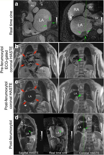
Fig. 8.

Pre-ferumoxytol ECG-gated HASTE and post-ferumoxytol HASTE CMR images without ECG-gating (3 T) belong to a 71-year-old female with dilated cardiomyopathy and bouts of sustained ventricular tachycardia (VT). FE-CMR was ordered to delineate a possible left atrial (LA) mass prior to undergoing VT ablation. Real-time cine FE-CMR images (panel a) demonstrate a large, sessile, immobile mass (green arrows) in the posterolateral wall of a severely dilated left atrium (red arrows). Compared to ECG-gated pre-ferumoxytol HASTE imaging (panel b), FE-HASTE CMR (panel c and d) achieved more complete and homogenous blood signal suppression, which enabled confident and definitive diagnosis of a left atrial mass (green arrows). The morphologic appearance of the left atrial mass on FE-HASTE CMR correlated well with findings on bright-blood real-time cine imaging (panel a; panel d, center image). Ao, aorta; LV, left ventricle; RV, right ventricle
