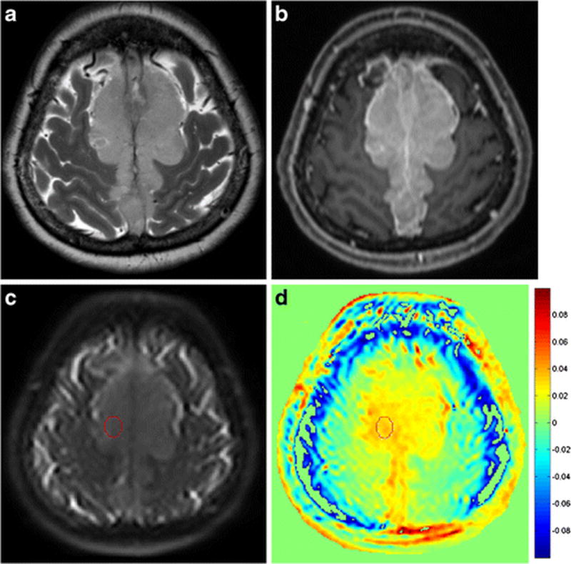
Figure 3.

A 57-year-old female patient with atypical meningioma exhibiting intermediate signal intensity on a T2-weighted image (a) and homogeneous enhancement on a contrast-enhanced T1-weighted image (b). A ROI was drawn on area which showed the highest signal within the tumour on a raw APT image (c) and then transferred to the processed APT-weighted image (d). The normalized magnetization transfer ratio asymmetry value within the enhancing tumour was 3.12 (c).
