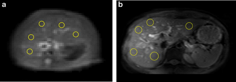
Fig. 1.

A&B. An example of placement of ROIs on a rat liver parenchyma region of T2 weighted image (A) and CEST image (B); C: An example of placement of ROIs on a human subject liver parenchyma region of CEST image. Artifacts and blood vessels were excluded.
