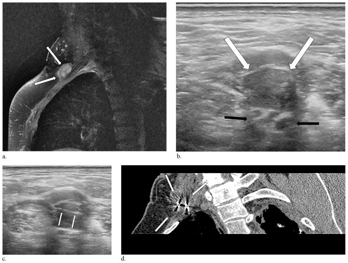
Figure 3.
A 54-year-old man presented with a 9-year history of upper extremity, lightning-like phantom sensations involving his lateral fingers and forearm. (a) Magnetic resonance imaging performed 2 years before the procedure demonstrates a 1.5 cm × 2.6 cm brachial plexus neuroma (arrows). (b) Ultrasound performed the day of the injection delineates the neuroma (white arrows) adjacent to the brachial plexus cords (black arrows). (c) Ultrasound guidance was employed in this case to place the cryoprobes (arrows) into the neuroma. (d) Reconstructed oblique CT image obtained 8 minutes into the first freeze cycle demonstrates 2 cryoprobes (stars) and the ablation zone engulfing the neuroma (arrows).

