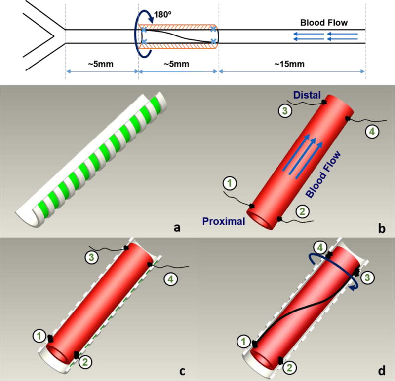
Figure 1. Schematics illustrating the twisting of rat carotid artery in vivo.

Top: location and dimension of the twisting segment. Bottom: surgical procedure: a) rigid semi-circular tubular sheath to support twisting; b) artery segment selected with 4 sutures attached to adventitia (2 at two sides of the vessel at the proximal and distal ends, respectively; c) Two sutures at the proximal end were sewn onto the sheath. d) The two sutures at the distal end were first swapped position to rotate (twist) the vessel 180° along its axis, and then sewn onto the sheath to hold the twist in the artery segment.
