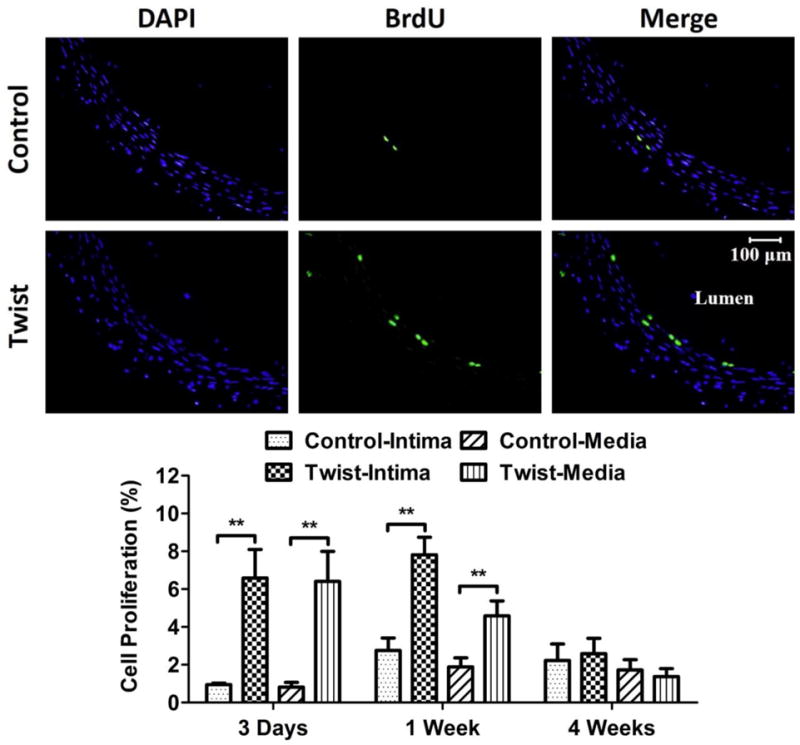
Figure 6. Twist induces cell proliferation in the arterial wall.

Photographs: Representative cross-sectional images of DAPI counter-staining and BrdU staining of the control and twisted arteries. Bargraphs: Comparison of cell proliferation ratio (mean ± SD, n=6) in the intima and media of arteries from the control and twist groups. ** p < 0.01 vs. controls.
