FIGURE 6:

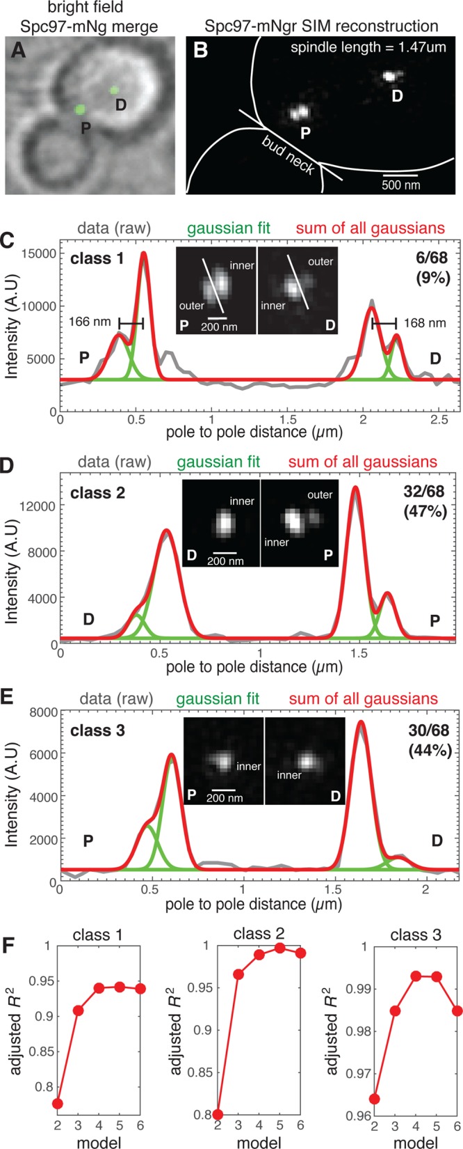
The γ-TuC protein Spc97 is present on the outer plaque of both SPBs of most metaphase spindles. (A) Spc97-mNG fusion protein localizes to both SPBs of metaphase spindles. (B) SIM reconstruction of Spc97-mNgr (spindle length = 1.47 µm) resolves densities representing the inner and outer plaques of SPBs that are proximal and distal to the bud neck. (C–E) The SIM reconstructions were used to classify 68 spindles with regard to visual detection of outer plaque densities; (C) both SPBs (class 1; 9%), (D) one SPB (class 2; 47%), or (E) neither SPB (class 3; 44%). For a representative spindle of each class, the fluorescence intensity (gray line) along the spindle axis of the SPBs shown, overlaid with the cumulative distribution (red) of the Gaussian mixture model for four individual peaks (green), with each peak representing distinct Spc97-mNgr densities. (F) The adjusted R2 for the Gaussian mixture model for peak numbers ranging from 2 to 6 for each class. The model fit improves with increasing peak number with goodness of fit increased at four peaks relative to two or three (densities for inner and outer plaques of both SPBs). This analysis reveals Spc97-mNgr outer plaque densities that are not visually resolved due to the axial resolution limit of SIM as well as a spindle axis that is frequently tilted relative to the focal plane.
