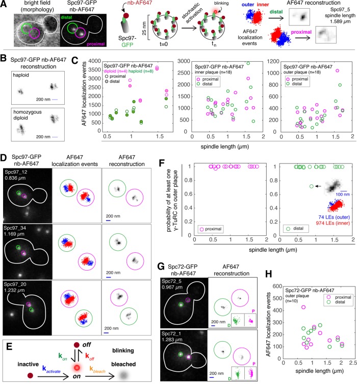
FIGURE 7:
Outer plaques of proximal and distal SPBs contain at least one γ-TuC complex. (A) SPB inner and outer plaques are not resolvable by conventional light microscopy with Alexa-647-conjugated nanobodies (GFP-trap; Chromotek) used to label Spc97-GFP. We applied dSTORM, a blinking-based superresolution method that can resolve the inner and outer plaques, with at most one blink per frame. For each blink, a single molecule is localized and this localization event can be represented as a point in space. A two-dimensional distribution of localization events representing the inner (red) and outer (blue) plaques of both SPBs is the starting point to generate a dSTORM reconstructed image by discretizing the space into 24-nm pixels. (B) Representative Spc97-GFP nb-AF647 reconstructions of inner and outer plaques were used to compare the inner plaque dimensions of SPBs in haploid and diploid cells. Bar in reconstruction is 200 nm. (C) Number of localization events for Spc97-GFP-AF647 for the inner plaques of diploids and haploids (left panel) and on the inner and outer plaques of haploids (middle and right panel). (D) Representative images (confocal, distribution of localization events and reconstruction) of SPBs in fixed cells, with Spc97-GFP labeled with nb-AF647. Sample name and spindle length is provided in the confocal image. Bar in reconstruction is 200 nm. (E) A simplified kinetic model of one active (blinking) and six inactive Spc97-GFP-AF647 molecules within a single γ-TuC complex. Inactive and bleached states correspond to the long-lived dark state. We assume that the fluorophore exits and enters the long-lived dark state once and only once during the acquisition time. (F) Probability of having at least one complete γ-TuC on the outer plaques of proximal and distal SPBs. With one exception (shown in inset), all cells in the data set (n = 18) are predicted to have least one γ−TuC on the outer plaque of both the proximal and distal SPBs. (G) Confocal image and AF647 reconstruction for Spc72-GFP detected with nb-AF647. Localization events for proximal and distal SPBs are shown as insets in the reconstruction image. (H) Number of localization events for Spc72-GFP-AF647 on the outer plaque.

