FIGURE 5:
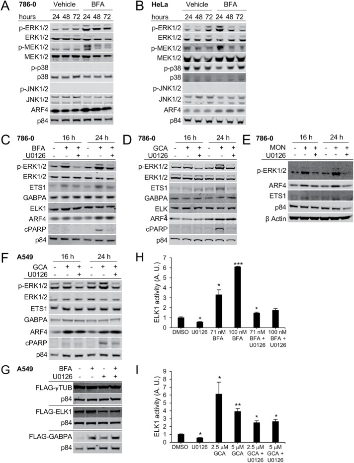
BFA treatment activates MEK1/2 and ERK1/2, but not p38 or JNK1/2 signaling. (A) 786-0 or (B) HeLa cells were treated with 40 nM BFA for the indicated duration. The indicated proteins were detected by Western blot. (C–E) 786-0 cells were treated for the indicated duration with 40 nM BFA (C), 1.75 µM GCA (D), or 500 nM MON in the presence or absence of the MEK inhibitor U0126, which was used at 10 μM. The specified proteins were detected by immunoblotting; cPARP: cleaved PARP. (A–E) Representative blots of one example out of three independent experiments are shown with the exception of E, where n = 1. (F) A549 cells were treated with 1.75 µM GCA in the presence or absence of the MEK inhibitor U0126 used at 10 μM for the indicated duration. (G) A549 cells stably expressing the indicated constructs were treated with 70 nM BFA in the presence or absence of 10 μM U0126 for 24 h. (H, I) ELK1 activity in response to 20-h treatment with the indicated concentrations of BFA (H) or GCA (I) in the absence or presence of 10 µM U0126 was measured using a dual-luciferase reporter assay. A representative example of two independent experiments (single compound treatment) is shown; the combinatorial treatment was done once. Three wells per condition were analyzed per experiment. A.U. indicates arbitrary units. Asterisks (*) represent significant differences between DMSO and treatment conditions: *, p < 0.05; **, p < 0.01; ***, p < 0.001.

